
Laptop Ultrasound Scanner(CE Approved)

Sally Yang
Contact person
Basic Information
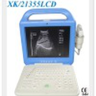
The digitallaptop Ultrasonic Diagnosis Equipment, is characterized by its clear image,stable performance and high resolution with adopting variable aperture,multi-segment dynamic focus,large dynamic band low-noise pre-amplifier, logarrthmic compression, TGC control, dynamic filter, edge enhancement and frame-related technologies etc. Standard configuration: Main unit 12″high resolution monitor R60 3.5MHz convex array probe USB connector Video out VGA socket Options High-frequency linear probe (5.0MHz, 7.5 MHz) Trans-vaginal probe (5.0MHz, 6.5 MHz) Trolley and portable type Technical Specifications: 1. Application: diagnosis of human liver, gallbladder, spleen, kidney, pancreas, heart, bladder, womb, urinary system intrauterine organ, pregnancy and the field of OB/GYN 2. Scanning depth (mm) ≥240 3. Detective Depth (mm) ≥190 4. Resolution (vertical direction) ≤1mm (depth ≤130mm) ≤2mm(130mm depth ≤170mm) Resolution (cross direction) ≤1mm (depth ≤130mm) ≤2mm(130mm≤depth ≤160mm) 5. Blind area (mm) ≤4 6. Imaging mode B, B+B, 4B, B+M, M, Zoom 7. Grey scale 256 8. Dynamic range ≥240 9. Cine loop 256-frame cine loop and 32-frame image storage 10.Electronic focusing dynamic receiving focusing 11. Frame correlation 4 segments frame correlation (B, B+B, 4B) 12. 7 kinds of colors (including of black and white) 13. Image turning left/right reverse, black/white reverse, up/down reverse 14. Measurement & calculation distance, perimeter, area, volume,pregnant weeks( BPD,GS,CRL,FL,AC,HC),Fetal weight,EDD and amniotic fluid etc. Heart rate and slope can be measured with Model M 15. Movie replay uninterrupted replay or step forward/backward replay for 256 images 16. Character input/display time, date, patient’s ID, age, sex, 17. Probe connector 2 18. 33 kinds of body marks 19. Image store Permanent 8 images 20. Voltage160V-260V, 50Hz±1Hz 21. Temperature scope +5°C +40°C 22. Relative scope ≤80% 23. 12.1 inch LCD monitor
Delivery terms and packaging
Packaging Detail: to be packed in cartons Delivery Detail: with in3 days after received payment
Port: chengdu/shenzhen
Payment term
Telegraphic transfer
Western Union

-
Payment Methods
We accept: